I came to work the other day to find this email waiting for me. I’m pleased to see that even when we get it wrong we sort of get it right.
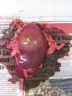
“This is a shot of the dissection of one of the porcine kidneys we sourced from you. As you can see it has a cyst. It was fascinating and the girls (and the staff) learnt so much. When it was dissected, it used urine, so we assumed there was a blockage – perhaps a stone. The girls handled and felt the difference between the healthy flesh and the unhealthy. I thought you’d be interested in our lesson.”
I’m not sure how we missed it when we were packing because we do check every organ that comes through, but in this case I’m glad we did.
Mr Vivi came back from a Chamber of Commerce talk the same day buzzing about this specky new machine they have at a nearby University. It allows the user to navigate and interact with all kinds of virtual environments – including body systems. I’m certain that it’s a fantastic, engaging and valuable educational tool but I’m just as certain that nothing is like experiencing the real thing in your hands.

Apr 10, 2014
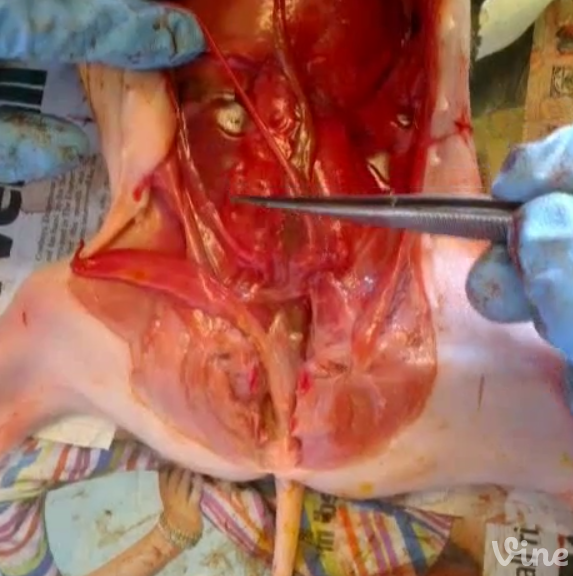
A quick look at the parts of the urinary system in a female stillborn piglet. You may need to click on the speaker icon to hear the voiceover by our lovely assistant Mr Vivi as he points out kidneys, ureter, bladder and urethra
— DissectionConnection (@missvivisection) November 20, 2013

We’ve gathered some new mammal dissection guides on Pinterest for you:
If you’ve found a good dissection guide that you’d like to share with others drop me a line with a link and I will be happy to post it there for you.
![]()
![]()