|
||||
|
|
||||
|
Every year at the end of June we get to go to ConQEST and we always have a ball.

This year we ran two workshops – a head dissection and a piglet dissection. One of the workshoppers, a lab tech from Emerald, took some great photos of the piglet dissection and has been kind enough to let me share them here.
Step 1 – peg out the beastie on a tray using rubber bands around each foot. Heather from Southern Biological showed me how to do this.
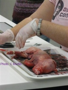
Step 2 – make a mid-sagittal incision in the skin
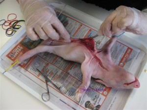
Step 3 – separate the skin from the muscle using a scalpel
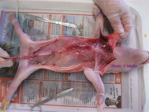
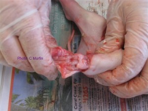
Step 4 – locate the diaphragm and identify the organs of the thoracic cavity
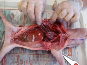
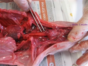
Step 5 – identify the organs of the abdominal cavity
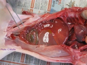
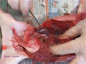
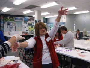
So, there you have it. A good time was had by all and then we went to lunch – which is always fabulous at ConQEST. See you there ‘in the flesh’ next year.
![]()
Hey Miss Vivi, just had 2 classes (Year 12) do the pig dissection. They loved it! Rated it their top dissection.
They have also done shark, toad and rat and the pig is by far the most popular. It was wonderful to see every boy engaged and taking part.
Thank you – from the Year 12 Biol students and teachers at BGS!!!
– Christina Jensen, Brisbane Grammar School
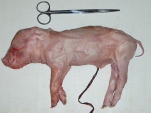
This little piggy is the latest and greatest in whole animal dissection specimens. They are either stillborn from large litters or smothered by the sow in the stall and come from a farm breeding pigs for meat. They are usually disposed of as farm waste but we are collecting them and diverting them from the waste stream for dissection. They are less smelly, cheaper, closer to human anatomy than a rat and aren’t being bred just to be euthanased for science, so each piglet used in the classroom represents a rat that hasn’t had to be put down.
Each piglet typically weighs 600-800g. The piglets are collected and frozen without any chemical preservatives which reduces your chemical exposure in the lab as well as eliminating a source of exposure to the kids in the classroom. It also makes the piglets safe to dispose of in landfill with other normal waste.
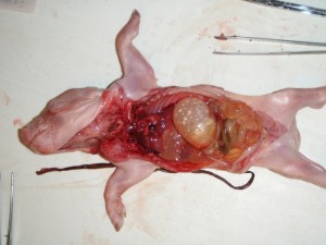
As they generally have not yet fed or only briefly suckled, the intestinal tract is pretty clean and there is little smell to the specimen. Each organ can easily be identified and removed for further exploration.
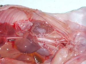
The detail in the circulatory system of the piglets is particularly amazing. You can see in the photo above the veins and arteries of the urogenital system clearly visible. Once the overlying organs were taken out, the spine was visible and a section could be removed to allow the spinal cord to be seen.
There is a lot more that can be explored with these specimens and we will be spending a lot of time working on them so we can develop some really good resources to allow you to get more bang for your buck. In the meantime keep an eye out for workshop announcements that will give you the chance to get your hands on one. We are expecting to be able to bring them to you at ConQEST 2012, if not before.
See you there,
![]()