Posted on

Top tip: spinal cord and brain dissection in piglets

Teaching science can be a tough gig.  Most of the time teachers feel that they are time poor and content rich.  Fitting everything required by the curriculum into a term can feel like herding cats into a bag.

Teachers know that kids want to explore science more than be taught it, but often it’s just not possible to let them take as much time as they need to really get as much learning out of an experiment as they would like.

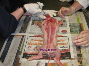
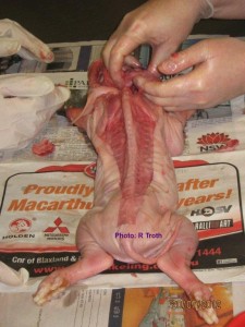
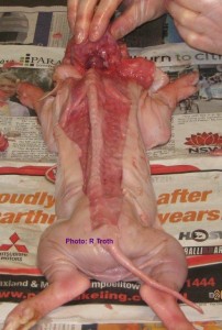
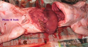
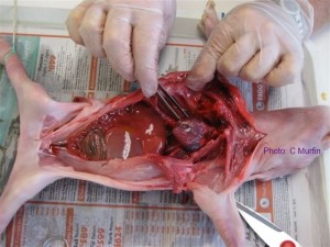
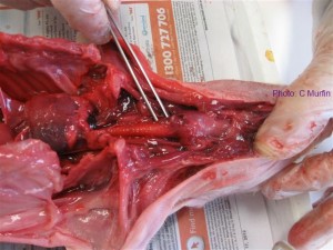
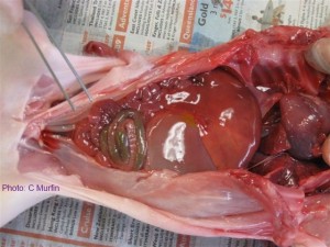
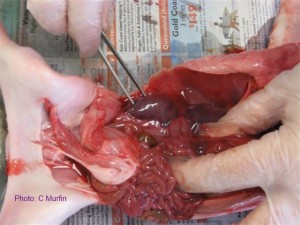
That’s why I’m over the moon when I hear that a class has had the time to really get more bang for their buck out of one of our specimens.  These photos have been sent by a labbie who bought them for a teacher wanting to teach about the brain and spinal cord.  We decided that piglets would be the best option to teach this and the kids were enjoying it so much that when they were finished looking at that they flipped them over and got into the abdominal organs as well.  Truly a case of learning about everything but the squeal.

The labbie says:

“They are an excellent class. It was great to see how involved they were during the prac. They really enjoyed it!
Because this dissection took place over just one period, I was requested to prepare the piglets prior to the prac, exposing the brain and the spine. … We couldn’t get hold of a Dremmel at the time so, used a hack saw to cut a cross in the skull then, cut the hole out with a pair of kitchen scissors.  He also suggested that a Dremmel would be perfect for this job. It’s been put on our list for purchases prior to the budget rollover.
I then prepared the others which made it quicker and easier for the students to then get to where the brain stem and the spinal cord meet since that was their main aim for the prac.
After that the piglets were dissected even further as you saw, to locate and expose all other organs in the stomach cavity etc
All of our Science staff were extremely impressed on the quality the specimens you sent.”
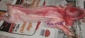
 

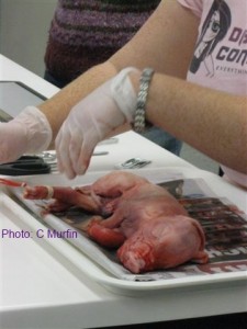
Spine and ribcage of the piglet
Piglet prepared for the class with spine and ribcage exposed
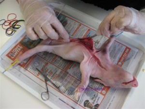
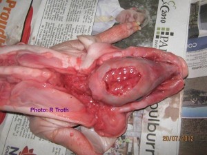
Brain of the piglet exposed
Base of the skull being opened
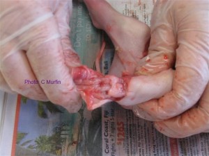
Brain stem of the piglet being examined
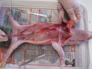
Spine of the piglet exposed
Abdominal organs in the piglet

Before we sent the piglets we had a bit of discussion by email about the best course of action for this prac. I still don’t have a Dremel and haven’t had a go at it, but this is what we suggested:

We believe that the best tool for opening the skull will be a small circular saw called a Dremel available at hardware stores. I was speaking to a very experienced biology teacher … that runs an extensive comparative anatomy program in his Yr 12 classes who agrees that they are the tool for the job. I am going to buy one and have a go myself. Do be careful because the skull will be slippery. You will use the Dremel to score the skull and then need a chisel and hammer to ease the skull open. There are some very good videos on YouTube that will show you what to do.

To expose the spinal cord at the back of the neck you will need to cut into the skin on either side of the spine at the base of the skull and lever the section away from the body and towards the tail. You will also be able to view the spinal cord and spinal nerves by opening the abdomen of the piglet and removing the organs to expose the spine. You will be able to see the spinal nerves entering/exiting the spinal cord cavity between the vertebrae – lift them with a probe. The vertebra will be soft enough to slice the top off with a scalpel and you will be able to view the spinal cord in-situ.

This website is an excellent resource for piglet dissections and the skull and spinal cord sections are very clearly described with photos. http://www.whitman.edu/content/virtualpig

Posted on

Top Tip: How to use small intestines in the classroom

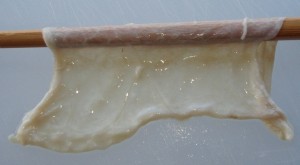
porcine small intestine
Porcine small intestine

I heard this from a lab tech from northern NSW at ConQEST.  She did such a good job with the small intestine in the science classes that now she’s asked to bring it into the hospitality classes as well.

She puts the specimen into an ice cream container and lets a little bit hang over the side.  Then she gets the kids to gather around and guess at what it is.  They can touch it at this stage but they can’t see what it looks like in the container.

After a while they get to see it all coiled up in the container and everyone gets around to working out that it’s a small intestine.  Then they start to wonder how long it is.  They’ve heard that these things can be metres long, so they are allowed to start extending it out.

They pull and they pull and they pull.  Eventually they are all standing holding this thing and then the labbie gets the tape measure out.  It’s around 6m long.

All the while these kids have been hearing about what the purpose of the small intestine is, where it fits in the jigsaw puzzle that is your abdominal cavity and what it connects to and so on.  They are fascinated by the thing and asking lots of really good questions.  Before they know it these kids can spout off all sorts of fun facts about their digestive system.  Teaching by stealth – I love it!

The specimen is tough enough to handle this over and over again.  You will store it quite happily in the fridge for years before you need a new one.  If you’re willing to you can sacrifice a little bit and make a wet mount to be viewed under the microscope.  Look out for the little ‘hairs’ along the edges – those are the remnants of all the circulatory system structures that bring oxygen to the small intestine and carry fluid away from it.

This is definitely a specimen that gives you a lot of bang for your buck.

Miss Vivi

Posted on

Piglet Dissection Workshop at ConQEST 2012

Every year at the end of June we get to go to ConQEST and we always have a ball.

Trade display at ConQEST 2012
The Dissection Connection trade display at ConQEST 2012

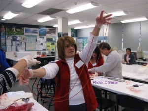
This year we ran two workshops – a head dissection and a piglet dissection.  One of the workshoppers, a lab tech from Emerald, took some great photos of the piglet dissection and has been kind enough to let me share them here.

Step 1 – peg out the beastie on a tray using rubber bands around each foot.  Heather from Southern Biological showed me how to do this.

Displaying the specimen using rubber bands
Piglet dissection: a rubber band around each foot and looped behind the dissection tray will hold the piglet still for dissection

Step 2 – make a mid-sagittal incision in the skin

Start with a mid-sagittal incision
Piglet dissection: making the mid-sagittal incision

Step 3 – separate the skin from the muscle using a scalpel

Piglet dissection: Exposing the muscles under the skin
Piglet dissection: once the skin has been peeled back the muscles are exposed for examination
Piglet dissection: exposed tarsals
Piglet dissection: the tarsals exposed

Step 4 – locate the diaphragm and identify the organs of the thoracic cavity

Piglet dissection: examining the organs in the thoracic cavity
Piglet dissection: examining the heart in the thoracic cavity
Piglet dissection: the trachea
Piglet dissection: an incision can be made in the trachea and the lungs can be inflated with a syringe

Step 5 – identify the organs of the abdominal cavity

Piglet dissection: organs of the abdominal cavity
Piglet dissection: the liver and intestine inside the abdominal cavity
Piglet dissection: kidney exposed in the abdominal cavity
Piglet dissection: intestine removed and kidney exposed
Piglet dissection: examining the length of the small intestine
Piglet dissection: the small intestine removed and stretched out to demonstrate its length

So, there you have it. A good time was had by all and then we went to lunch – which is always fabulous at ConQEST.  See you there ‘in the flesh’ next year.
Miss Vivi

Posted on 2 Comments

Free posters for the classroom: hand washing and glove removal techniques

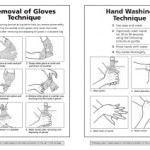
hand washing and glove removal posters
Free posters for the classroom: hand washing and glove removal technique
germ farm poster
Poster for the classroom: scrub 'em! poster from www.1st-in-handwashing.com

I think sometimes we forget that we aren’t born knowing how to do things ‘the right way’. I’ve been conscious of washing my hands properly and taking my gloves off without cross contaminating other surfaces for so long that I don’t even realise I’m doing it anymore. I remember being in the ladies at work one day washing my hands and a science teacher was standing next to me. She looked over at what I was doing and said:
“You wash your hands like a lab tech.”

I looked down and I was soaping between my fingers and I had suds right up around my wrists.

I’ve had these hand washing and glove removal posters for a long time and I’ve always put a copy up next to the sink in the classrooms where I’m working.  I can’t find a link to them anywhere now, but I do know that they were originally shared by some very generous labbie on a lab tech discussion list quite a few years ago.  I’ve also kept a laminated copy of each to put out with dissection equipment and I’ve always made a point of  getting around to each group in the classroom and showing the kids how to take their gloves off properly.  Click on the image to download a copy of them for yourself.

The “Scrub ’em!” poster is available from 1st-in-handwashing and is a bright, colourful reminder to wash your hands that appeals to kids so it’s good for toilets and around tuckshops.  Click on the image to see a bigger version.

Miss Vivi