Posted on

Piglet Dissection Workshop at ConQEST 2012

Every year at the end of June we get to go to ConQEST and we always have a ball.

Trade display at ConQEST 2012
The Dissection Connection trade display at ConQEST 2012

This year we ran two workshops – a head dissection and a piglet dissection.  One of the workshoppers, a lab tech from Emerald, took some great photos of the piglet dissection and has been kind enough to let me share them here.

Step 1 – peg out the beastie on a tray using rubber bands around each foot.  Heather from Southern Biological showed me how to do this.
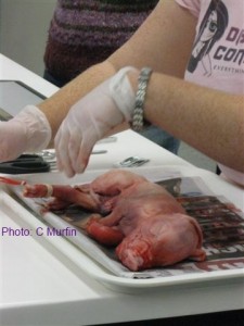
Displaying the specimen using rubber bands
Piglet dissection: a rubber band around each foot and looped behind the dissection tray will hold the piglet still for dissection

Step 2 – make a mid-sagittal incision in the skin
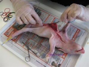
Start with a mid-sagittal incision
Piglet dissection: making the mid-sagittal incision

Step 3 – separate the skin from the muscle using a scalpel
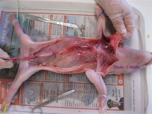
Piglet dissection: Exposing the muscles under the skin
Piglet dissection: once the skin has been peeled back the muscles are exposed for examination
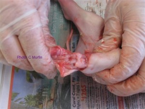
Piglet dissection: exposed tarsals
Piglet dissection: the tarsals exposed

Step 4 – locate the diaphragm and identify the organs of the thoracic cavity
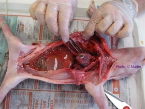
Piglet dissection: examining the organs in the thoracic cavity
Piglet dissection: examining the heart in the thoracic cavity
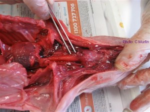
Piglet dissection: the trachea
Piglet dissection: an incision can be made in the trachea and the lungs can be inflated with a syringe

Step 5 – identify the organs of the abdominal cavity
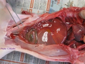
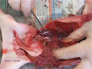
Piglet dissection: organs of the abdominal cavity
Piglet dissection: the liver and intestine inside the abdominal cavity
Piglet dissection: kidney exposed in the abdominal cavity
Piglet dissection: intestine removed and kidney exposed
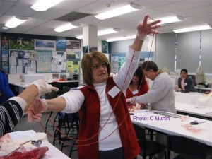
Piglet dissection: examining the length of the small intestine
Piglet dissection: the small intestine removed and stretched out to demonstrate its length

So, there you have it. A good time was had by all and then we went to lunch – which is always fabulous at ConQEST.  See you there ‘in the flesh’ next year.
Miss Vivi

Posted on

Top Tip: keeping slides warm for viewing semen motility

Now that the weather has cooled down it’s going to take a bit more to keep semen samples warm enough to view motility.  Live semen is sensitive to thermal shock and the glassware will need to be warmed up in a water bath to at least 35C before you introduce the sample.

A top tip from the supplier to keep the slide warm and slow down the rate of cooling of the glass:

  1. cut a piece of cardboard the same size as a microscope slide,
  2. cut a hole in it the same size and position as the cavity to allow the light through,
  3. mount the cardboard on the microscope stage,
  4. mount the warmed slide on top of the cardboard.

When you order the live semen sample I will send you an info sheet so you are prepared before it arrives, so don’t panic if you’re not sure what I’m talking about here.

And if you’re wondering whether or not to order the sample in the first place, why not have a look at what a labbie had to say about how they managed it and what the kids got out of it?


Posted on

Top dissection specimen as voted by students

Hey Miss Vivi, just had 2 classes (Year 12) do the pig dissection. They loved it! Rated it their top dissection.

They have also done shark, toad and rat and the pig is by far the most popular. It was wonderful to see every boy engaged and taking part.
Thank you – from the Year 12 Biol students and teachers at BGS!!!

– Christina Jensen, Brisbane Grammar School